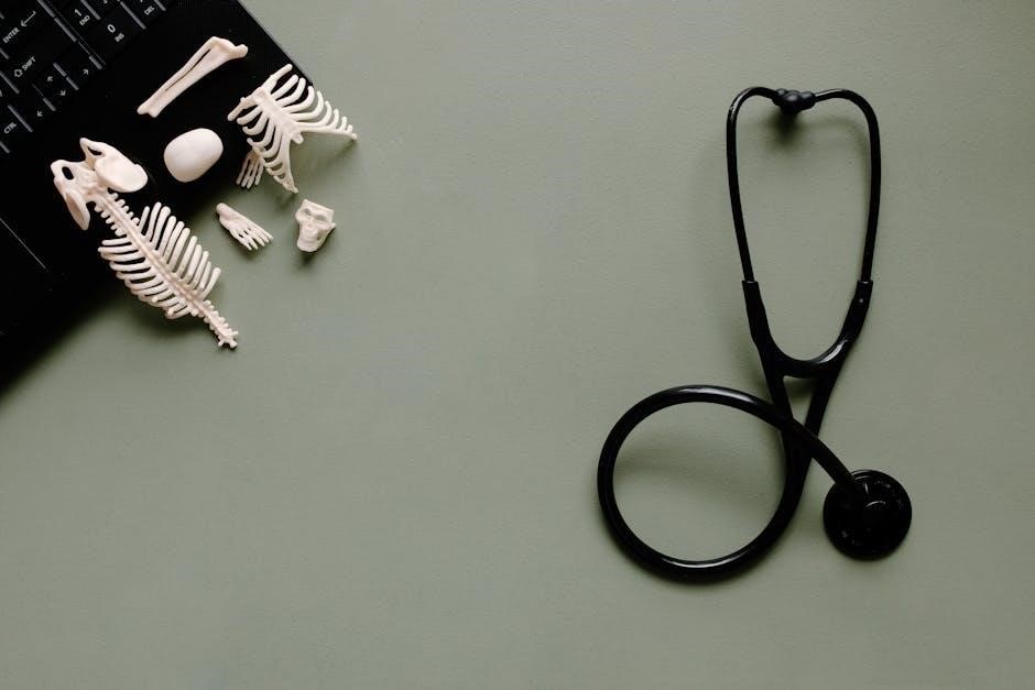
Welcome to the laboratory manual for human anatomy and physiology, a comprehensive guide designed to enhance your understanding of the human body through hands-on exploration. This manual provides structured exercises, safety protocols, and essential techniques to navigate the complexities of anatomical and physiological studies effectively.
Overview of the Laboratory Manual
This laboratory manual serves as a detailed guide for exploring human anatomy and physiology through practical exercises and investigations; It covers essential topics like histology, skeletal and muscular systems, and dissection techniques. The manual emphasizes safety, proper use of equipment, and hands-on activities to reinforce theoretical knowledge, providing a comprehensive learning experience for students in anatomy and physiology courses.
Importance of Laboratory Studies in Anatomy & Physiology
Laboratory studies are crucial for understanding the intricate structures and functions of the human body. Hands-on experiences, such as dissections and microscopy, enhance visual and tactile learning, reinforcing theoretical concepts. These practical exercises develop critical thinking, observation, and analytical skills, essential for careers in healthcare and scientific research, while fostering a deeper appreciation of human anatomy and physiology.
Essential Laboratory Tools and Equipment
Laboratory tools like microscopes, dissection kits, and measuring instruments are vital for anatomy and physiology studies, enabling precise observations, specimen handling, and accurate data collection.
Microscopes and Their Use in Anatomy & Physiology
Microscopes are essential tools for examining microscopic structures in anatomy and physiology. They enable detailed observation of cells, tissues, and organisms. Compound microscopes are commonly used for viewing histological slides, while stereo microscopes are ideal for studying three-dimensional specimens. Proper use involves adjusting focus, illumination, and magnification to clearly visualize specimens, aiding in understanding cellular and tissue structures.
Dissection Tools and Their Functions
Dissection tools, such as scalpels, forceps, and dissecting scissors, are crucial for carefully examining anatomical structures. Scalpels are used for precise cutting, while forceps aid in gripping tissues. Dissecting scissors help in separating layers without causing damage. These tools enable students to explore the internal organization of specimens, fostering a deeper understanding of anatomical relationships and functional structures.

Safety Protocols in the Anatomy & Physiology Lab
Adherence to safety guidelines is crucial in the anatomy and physiology lab. Proper use of PPE, safe handling of chemicals, and disposal of biological waste are essential. Ensure equipment is used correctly, and emergency procedures are understood to maintain a safe learning environment.
Handling Biological Specimens Safely
Handling biological specimens requires careful attention to safety protocols. Always wear appropriate PPE, including gloves and goggles, to prevent exposure. Ensure proper disposal of biological waste in designated containers. Clean and decontaminate equipment after use, and avoid eating or drinking in the lab. Adhere to all safety guidelines to minimize risks and maintain a safe environment for everyone.
Proper Use of Personal Protective Equipment (PPE)
Proper use of PPE is essential for protecting against biological and chemical hazards in the lab. Always wear gloves to prevent skin contact with specimens. Lab coats and closed-toe shoes shield clothing and feet from potential spills. Goggles protect eyes from splashes. Ensure all PPE is worn correctly and removed before leaving the lab area to maintain safety and hygiene standards consistently.
Histology: The Study of Tissues
Histology involves examining tissues under a microscope to understand their structure and function. This field is crucial for diagnosing diseases and studying cellular organization in various organisms.
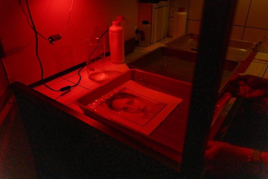
Preparation of Histological Slides
Preparing histological slides involves sectioning tissues, staining, and mounting them for microscopic examination. Sections are cut using microtomes, typically 4-5 µm thick. Staining enhances contrast, with hematoxylin and eosin (H&E) being common. Proper fixation, dehydration, and embedding ensure tissue preservation. Accurate preparation is critical for clear visualization and accurate tissue analysis under a microscope.
Identifying Different Types of Tissues Under a Microscope
Under a microscope, tissues are identified by their cellular arrangement and specialized features. Epithelial tissues appear as tightly packed cells, while connective tissues show a matrix with scattered cells. Muscle tissues exhibit striations, and nervous tissues display elongated neurons with dendrites. Staining techniques like H&E enhance visibility, allowing for precise differentiation and study of tissue structures and functions.
The Skeletal System: Structure and Function
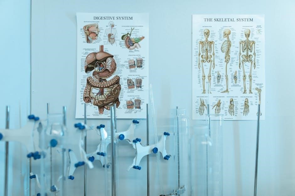
The skeletal system provides structural support, protects vital organs, and enables movement. Comprising bones and cartilage, it facilitates mobility through joints and supports overall body weight and function.
Identifying Bones and Their Landmarks
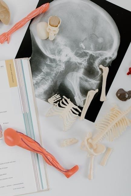
Accurate identification of bones and their landmarks is crucial for understanding skeletal anatomy. Use diagrams and anatomical terms to locate key features such as foramina, fossae, and epicondyles. Compare bones to recognize variations in shape and function, ensuring precise identification during lab exercises and dissections.
Lab Activities for Understanding Joint Movements
Engage in hands-on activities to explore joint movements, including flexion, extension, and rotation. Use skeletal models to visualize joint types and their functions. Measure range of motion and observe how muscles and ligaments influence movement. Palpation exercises help identify joint structures and their roles in mobility, enhancing your understanding of joint mechanics and their significance in human anatomy.
The Muscular System: Structure and Function
The muscular system consists of skeletal, smooth, and cardiac muscles, enabling movement, maintaining posture, and supporting bodily functions. Its structure and function are vital for human physiology.
Identifying Major Muscle Groups
The muscular system is composed of over 600 muscles, grouped into major categories such as upper limb, lower limb, abdominal, and back muscles. Key groups include the pectoralis major, deltoids, quadriceps, hamstrings, and latissimus dorsi. Understanding these groups is essential for comprehending movement and anatomical functions, aiding in both clinical applications and laboratory studies.
Lab Activities for Understanding Muscle Actions
Lab activities focus on identifying muscle functions through palpation, contractions, and movements. Students explore how muscles interact during actions like flexion, extension, and rotation. Practical exercises include observing muscle contractions in peer groups and using models to visualize muscle mechanics. These hands-on approaches enhance understanding of muscle physiology and their roles in movement, fostering a deeper connection between anatomy and function.
Dissection and Exploration of Human Cadavers
Dissection and exploration of human cadavers provide hands-on experience, offering deep insights into anatomical structures and their relationships. This lab work is crucial for understanding human physiology and preparing future healthcare professionals.
Step-by-Step Dissection Techniques
Mastering step-by-step dissection techniques involves careful preparation, precision, and adherence to anatomical guidelines. Start with proper instrument handling and systematic layer-by-layer exposure. Identify key structures, document findings, and maintain a clean workspace. These skills enhance anatomical understanding and promote safe, respectful exploration of cadavers, fostering a deeper appreciation of human anatomy and its functional complexities.
Identifying Organs and Their Locations
Identifying organs and their locations requires careful observation and understanding of anatomical landmarks. Major organs like the heart, lungs, liver, and kidneys are located in specific cavities. The thoracic cavity houses the heart and lungs, while the abdominal cavity contains the liver, stomach, and kidneys. Use dissection guides and anatomical charts to accurately locate and document each organ’s position and relationships with surrounding structures.
Microscopy and Staining Techniques
Microscopy and staining techniques are essential for examining cellular structures in anatomy and physiology. These methods involve preparing samples, applying stains, and using microscopes for clear visualization and accurate analysis.
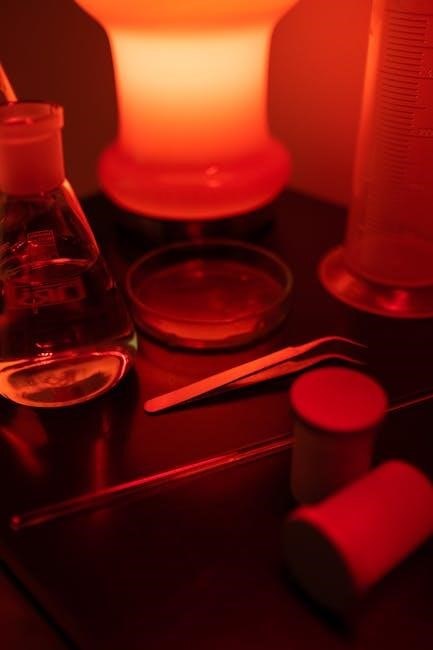
Preparing and Staining Tissue Samples
Preparing and staining tissue samples involves sectioning, fixation, and applying specific dyes to enhance visibility under a microscope. Fixatives preserve tissue structure, while stains like hematoxylin and eosin (H&E) differentiate cellular components. Proper techniques ensure clear visualization of microscopic structures, aiding in accurate anatomical and physiological analysis. This process is crucial for detailed tissue study in laboratory settings.
Interpreting Microscopic Structures
Interpreting microscopic structures requires adjusting the microscope for clarity and recognizing patterns in stained tissue samples. Different stains highlight specific cellular features, aiding in identifying tissues like epithelial or connective. Proper illumination and focus ensure detailed observation, enabling accurate analysis of anatomical structures for educational or diagnostic purposes.
Lab Activities for the Digestive System
Lab activities for the digestive system involve exploring the anatomy of the digestive tract and simulating processes like digestion and absorption. These exercises connect structure to function, enhancing understanding of how nutrients are processed in the body.
Understanding the Anatomy of the Digestive Tract
The digestive tract consists of the mouth, esophagus, stomach, small intestine, and large intestine. Each section specializes in mechanical and chemical digestion, absorption, or elimination of waste. Lab exercises focus on identifying these structures and their functions, emphasizing how they work together to process nutrients effectively.
Lab Exercises on Digestive Processes
Lab exercises simulate digestive processes, including ingestion, mechanical digestion, and chemical digestion. Students observe enzyme activity, nutrient absorption, and waste elimination. Practical activities involve testing enzyme effectiveness on food samples and analyzing digestion stages under a microscope, reinforcing understanding of the digestive system’s functionality and its biological importance.
Cellular and Tissue Studies
Cellular and tissue studies involve examining microscopic structures to understand cell functions and tissue organization, crucial for anatomy and physiology education and research.
Observing Cellular Structures Under a Microscope
Observing cellular structures under a microscope involves preparing slides, focusing the lens, and adjusting magnification to visualize cell membranes, nuclei, and organelles. Proper technique ensures clear images for accurate analysis.
Understanding Tissue Regeneration and Repair
Tissue regeneration and repair involve the restoration of damaged or lost cells, essential for maintaining bodily functions. This process varies across epithelial, connective, and muscular tissues. Factors like age and disease can influence regenerative capacity. Laboratory studies, including histological examinations, provide insights into these mechanisms, aiding in the development of therapeutic strategies for tissue engineering and regenerative medicine.
Practical Applications of Anatomy & Physiology Knowledge
Anatomy and physiology knowledge is crucial in clinical settings, sports, and fitness, enabling precise diagnoses, effective treatments, and personalized training plans. Lab findings often guide real-world applications.
Case Studies in Clinical Anatomy
Case studies in clinical anatomy provide real-world insights into human health and disease. By analyzing patient histories, symptoms, and diagnoses, students connect anatomical knowledge to clinical scenarios. These studies enhance understanding of how structural abnormalities impact function, aiding in precise treatment planning and fostering critical thinking in medical practice and research.
Connecting Lab Findings to Real-World Scenarios
Lab findings are crucial for translating anatomical and physiological knowledge into practical medical applications. By connecting these discoveries to real-world scenarios, students can better understand disease mechanisms, diagnostic techniques, and treatment strategies. This approach bridges the gap between theoretical learning and clinical practices, enhancing the ability to apply scientific knowledge to patient care and healthcare solutions effectively.